
嘉峪检测网 2025-06-16 20:31
导读:本研究旨在探究骨粉尺寸和消化时间对人骨组织来源 ECM 水凝胶特性的关键影响,为开发有效的骨修复生物材料提供 ECM 理化特性方面的见解。
去细胞组织因保留天然细胞外基质(ECM)的复杂成分和结构,在组织再生方面有巨大潜力。我们通过调整人骨ECM来源的ECM水凝胶的骨粉大小和酶消化时间来控制其理化及生物学特性。减小骨粉尺寸、延长消化时间可提高水凝胶的蛋白浓度、蛋白多样性和凝胶强度。HBMSCs在骨粉水凝胶上培养后,成骨分化增强,证实这些水凝胶是骨再生的生物活性材料。
01研究内容简介
1、引言
组织工程利用细胞、生物活性分子和生物材料修复受损组织,具有广阔的应用前景。生物材料在组织工程中至关重要,它能够提供微环境,并输送能促进再生组织中细胞功能的分子。诸多策略聚焦于开发模仿天然细胞外基质(ECM)结构和成分的生物材料。天然聚合物水凝胶,如胶原蛋白、海藻酸盐、壳聚糖、透明质酸和明胶,与 ECM 成分相似,且能形成纤维和多孔微观结构,调节细胞的黏附、增殖和分化等行为。但复制 ECM 中复杂的结构和功能蛋白混合物,包括糖胺聚糖、胶原蛋白、蛋白聚糖和生长因子,至今仍颇具挑战。
组织脱细胞化成为制备和应用生物材料的有效方法。去除天然组织中的细胞成分后,可保留 ECM 的复杂组成和结构,还能降低免疫原性,减少炎症反应和疾病传播。事实上,脱细胞 ECM 已被证实能促进细胞行为和组织重塑。目前,动物脱细胞基质已用于多种临床产品,如三维植入支架、二维薄片和粉末。最近,脱细胞 ECM 还被制成水凝胶。ECM 水凝胶主要由脱细胞 ECM 溶解或消化的蛋白质组成,能在生理温度和 pH 值下形成凝胶,可通过注射用于缺损部位,比 ECM 粉末分布更均匀。最常用的制备方法是用胃蛋白酶溶解脱细胞组织粉末,该酶能溶解 ECM 中的多种蛋白质,多项研究表明,基于 ECM 蛋白质的水凝胶保留了一定生物活性,能改善细胞增殖、分化和组织修复,自然也应用于组织工程和再生医学。
目前,关于牛和猪骨组织来源的脱细胞 ECM 水凝胶用于骨组织再生的研究有限,这些水凝胶已被证实具有增强细胞功能和促进骨再生的能力。本研究探索利用人供体骨移植材料制备人 ECM 凝胶,以替代动物组织来源的 ECM 水凝胶。无论组织来源如何,ECM 水凝胶都是去除细胞成分、免疫原性低的天然 ECM 材料。人体组织虽不易获取,但免疫原性最低,异体移植是治疗的金标准。动物组织来源的 ECM 可能因某些动物蛋白成分引发免疫反应,如牛胶原蛋白、蛋白聚糖连接蛋白、聚糖抗原、猪内源性逆转录病毒基因和 α - gal 表位。
在制备猪骨来源的 ECM 水凝胶时,脱矿脱细胞骨粉(<40 μm)在胃蛋白酶溶液中处理 1 小时。类似地,牛骨粉来源的 ECM 水凝胶需在胃蛋白酶溶液中消化 96 小时。然而,组织大小和消化时间对 ECM 水凝胶的蛋白质浓度、凝胶化、流变学和生物学特性的影响尚不明确。关键是,尚无针对人骨组织 ECM 水凝胶制备参数的研究。这对减少批次间差异(尤其是骨粉尺寸范围广的情况),以及通过优化 ECM 蛋白质比例最大化其再生功能至关重要。本研究专注于脱矿脱细胞人骨粉细胞外基质来源的水凝胶,与传统脱矿骨粉不同。
因此,本研究考察了颗粒大小和消化时间这两个制备人骨 ECM 水凝胶的关键参数,对其理化和生物学特性的影响,以及在骨组织修复中的应用潜力。人小梁骨组织取自骨科择期手术的人股骨头,并制备了不同尺寸(45 – 250 μm、250 – 1000 μm 和 1000 – 2000 μm)的脱矿人骨粉。这些骨粉经胃蛋白酶处理 3、5 或 7 天。基于骨粉尺寸和消化时间,评估所得 ECM 水凝胶的凝胶化和流变学特性,用扫描电子显微镜和显微计算机断层扫描检查水凝胶微观结构,通过测量细胞和蛋白质浓度评估蛋白质消化效率,用质谱法评估水凝胶的蛋白质组学特征,最后在人 ECM 水凝胶上检测人骨髓基质干细胞的黏附、铺展、迁移、增殖和分化,以确定其生物学功能。
总之,本研究旨在探究骨粉尺寸和消化时间对人骨组织来源 ECM 水凝胶特性的关键影响,为开发有效的骨修复生物材料提供 ECM 理化特性方面的见解。
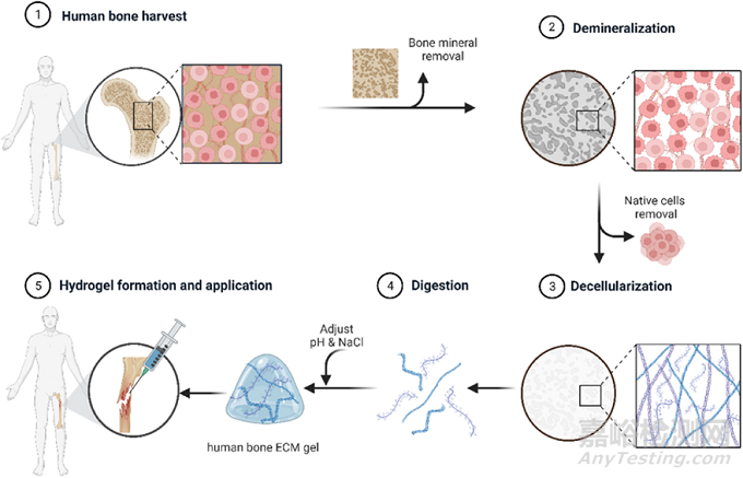
Fig. 1. Schematic illustration of ECM hydrogel derived from demineralized and decellularized human bone.Human trabecular bones were demineralized and decellularised. The digested extracellular matrix proteins were crosslinked, forming a gel to apply to a defect.
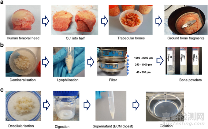
Fig. 2. Preparation process of ECM hydrogel derived from demineralized and decellularized human bone. (a) Human femoral heads from haematologically normal patients, both female and male, aged 65 ± 12 years old, were cut in half, and the trabecular bones collected and fragmented. (b) The fragments underwent demineralization in 0.5N HCl for 24 h, followed by lyophilization. The lyophilised powders were then sieved through stainless steel sieves to obtain powders in the ranges of 45–250 μm, 250–1000 μm, and 1000–2000 μm. (c) After decellularization in a Trypsin/EDTA solution, the powders were digested in a pepsin solution for 3, 5, and 7 days, respectively. The supernatant from the digestion solution was mixed with 0.1N NaOH, 10 × PBS, and 1 × PBS and incubated at 37 ◦C for 1 h to induce gelation.
2.结果与讨论
在生物材料和组织再生医学领域,复制天然细胞外基质(ECM)的特性一直备受关注。本研究旨在开发来源于人骨ECM的水凝胶,以应用于骨科修复领域。我们假设ECM骨粉颗粒的大小和ECM消化的持续时间对ECM衍生蛋白质的质量和数量有重要影响,进而影响所得水凝胶的流变学和物理化学性质。通过考察人基质细胞功能,我们探讨了人骨ECM水凝胶在促进骨组织修复方面的潜力。(图2)

Fig. 3. Gelation and rheological characterizations of ECM hydrogels from various powder sizes and digestion time. (a) Turbidimetric gelation kinetics of ECM hydrogels. pH-neutralized ECM pepsin digests were added to the wells of a pre-warmed 96-well plate (37 ◦ C), and the absorbance at 405 nm was measured at 3-min intervals (n = 4). The values were normalized between 0 (the initial absorbance) and 1 (the maximum absorbance). (b) Photograph images of each gel after pH neutralization in inverted tubes before and after incubation at 37 ◦C. The red line indicates the initial volume of the gel before inversion. (c) S (speed of gelation), tlag (time to start gelation), and t1/2 (time to reach 50 % maximum turbidity) were calculated based on turbidimetric curves. (d and e) Storage modulus (G′, open marks) and loss modulus (G″, closed marks) were monitored as hydrogels underwent an amplitude sweep of 0.1–100 % strain at a constant angular frequency. Data represent means ± standard deviation for n = 3. (f) Fourier transform infrared spectroscopy (FTIR) spectra of ECM digest compared with collagen with and without ECM buffer were analyzed. The spectrometer’s detection range was 399–4000 cm-1, and the data were measured with an interval of 0.96 cm–1 at room temperature. Statistical significance was determined using a two-way ANOVA test with Tukey’s multiple comparisons test (*p < 0.05 **p < 0.005 ***p < 0.0005****). The † symbol in-dicates a significant difference within the groups at the same digestion time). Data represent mean ± SD, N = 4.
3.1 骨粉大小和消化时间对水凝胶性质的影响
我们制备了多种类型的人骨ECM水凝胶,并研究了骨粉大小和消化时间对水凝胶凝胶化和流变性质的影响。通过浊度法凝胶化动力学分析,我们发现所有水凝胶的凝胶化动力学均呈现S型模式。特别地,来自较小骨粉(45-250 μm)的水凝胶,无论消化时间如何,都表现出比来自较大骨粉(250-1000 μm和1000-2000 μm)的水凝胶更陡峭的斜率和更短的凝胶化时间(tlag和t1/2)。具体来说,45-D3/D5/D7水凝胶在37℃孵育后约7分钟内即发生凝胶化,而250-D3/D5/D7和1000-D3/D5/D7水凝胶的凝胶化时间分别为16分钟和20分钟。 此外,我们还评估了水凝胶的流变特性。结果显示,来自较小骨粉(45-250 μm)的水凝胶,特别是经过5天和7天消化的水凝胶,其储能模量(G')显著高于来自较大骨粉的水凝胶。这表明较小骨粉尺寸和较长消化时间能够溶解出更多种类的蛋白质,从而生成具有更高机械强度的水凝胶。 为了测试水凝胶的稳定性和体外降解情况,我们评估了水凝胶在PBS和胶原酶-PBS中的蛋白质释放量。结果显示,在PBS中孵育后,约20-25%的蛋白质从ECM水凝胶中释放,且该比例在较长时间内保持稳定。然而,在加入胶原酶-PBS溶液后,所有水凝胶中释放的蛋白质浓度迅速增加。值得注意的是,来自较小骨粉样本的水凝胶表现出较慢的蛋白质释放曲线,表明其降解速度较慢。(图3)
3.2 ECM水凝胶的凝胶化机制
我们研究了ECM水凝胶的凝胶化机制,并发现其与胶原蛋白提取物的凝胶化过程相似。通过中和ECM消化物的pH值和盐浓度,并在37℃下孵育,可以诱导ECM水凝胶的形成。为了探究凝胶化过程中胶原蛋白的贡献,我们使用傅里叶变换红外光谱(FTIR)分析了中和ECM缓冲液对胶原蛋白和ECM消化前凝胶化学结构的影响。结果显示,ECM缓冲液中的NaCl和NaOH在胶原蛋白和ECM水凝胶的凝胶化过程中起着重要作用,它们通过形成胶原蛋白多肽链之间的连接来促进凝胶化。(图3)
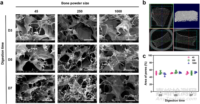
Fig. 4. Microstructure analysis of human bone ECM hydrogels using scanning electron microscope (SEM) and micro-computed tomography (μ-CT). (a) SEM images of cross-sections of lyophilised ECM hydrogels were observed by SEM (scale bar = 300 μm), showing the presence of porous and fibrous structures in all types of ECM hydrogels. (b) 3-dimensional visualization of the ECM hydrogel structure using μ-CT demonstrates the porous nature of the hydrogels. (c) Measurement of pore sizes in ECM hydrogels using ImageJ software reveals no significant differences between the hydrogels (n = 3). Statistical significance was determined using a two-way ANOVA test with Tukey’s multiple comparisons test (*p < 0.05 **p < 0.005 ***p < 0.0005****). Data represent mean ± SD, N = 4.
3.3 水凝胶的微观结构
我们检查了水凝胶的横截面和三维结构,发现所有水凝胶均表现出多孔和纤维状结构。纤维广泛分布在孔隙中,这些孔隙相互连接,为细胞提供了良好的生长环境。(图4)
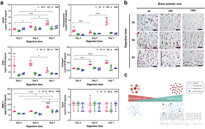
Fig. 5. Concentrations of DNA, total proteins, sGAG, collagen, BMP-2 and VEGF in ECM digests and histological staining images of ECM hydrogels. (a) Significantly high concentrations of proteins, including sGAG and BMP-2, were detected in the 45–250 μm powder and 7-day digested ECM digest, compared to other ECM digests (n = 4). Statistical significance was determined using a two-way ANOVA test with Tukey’s multiple comparisons test (*p < 0.05 **p < 0.005 ***p <0.0005****). The † symbol indicates a significant difference within the groups at the same digestion time). Data represent mean ± SD, N = 4. (b) ECM hydrogels were stained with haematoxylin and Alcian blue/Sirius red (scale bar = 100 μm). (c) Schematic illustration of the effects of bone powder size and digestion time on digesting ECM proteins.
3.4 ECM消化物中的细胞和蛋白质含量
我们量化了ECM消化物中DNA和蛋白质的浓度,并发现总蛋白质、sGAG和胶原蛋白的浓度随着骨粉尺寸的减小和消化时间的增加而显著增加。此外,我们还评估了ECM去细胞化的效果,并发现较小骨粉尺寸中的DNA浓度显著高于较大骨粉,但随着消化时间的增加,DNA浓度逐渐降低。这表明骨粉大小和消化时间对于从细胞外基质中消除细胞至关重要。 为了可视化水凝胶中蛋白质的分布,我们使用了特定染料进行染色。结果显示,水凝胶在胶原蛋白和GAG方面表现出显著的染色,而细胞核染色可忽略不计。这表明水凝胶中含有丰富的胶原蛋白和GAG,这对于促进细胞附着和生长至关重要。(图5)
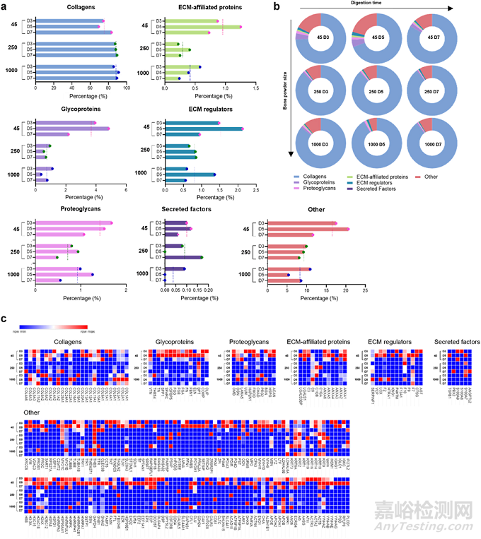
Fig. 6. Comparison of the total matrisome subcategories of proteins in human bone ECM digests. (a and b) illustrate the percentages of proteins categorized under different matrisome subcategories in ECM digests. ECM digests derived from various sizes of bone powders after 5 days of digestion exhibit a significant impact on the protein proportions compared to those from shorter or longer incubation times. The dashed lines in (a) represent average percentages from the group. (c) The details and proportions of the proteins are listed in the heatmap.
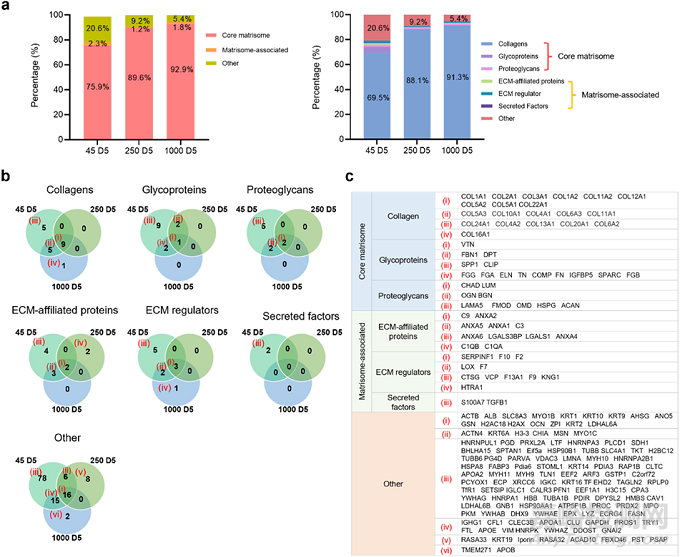
Fig. 7. Total matrisome subcategories of proteins in human bone ECM digests from 45-D5, 250-D5, and 1000-D5. (a) presents the percentages of categorized proteins from 45-D5, 250-D5 and 1000-D5. (b) shows a Venn diagram depicting the percentage of matrisome subcategories of proteins in various sizes of bone powders after 5 days of digestion. Smaller bone powder exhibits a lower presence of collagens but a higher presence of other protein types. (c) The details of the proteins are listed in the table.
3.5蛋白质组学分析
为了更深入地了解ECM水凝胶中蛋白质的组成,我们进行了蛋白质组学分析。结果显示,在ECM消化后,我们最多鉴定出了246种蛋白质,包括核心基质蛋白质、基质相关蛋白质和非基质蛋白质。我们发现,骨粉大小和消化时间对ECM消化物中分类蛋白质的比例有重要影响。具体来说,较小骨粉尺寸和较长消化时间能够溶解出更多种类的蛋白质,且这些蛋白质在ECM水凝胶中的比例也更高。 此外,我们还发现,在较小骨粉尺寸和中等消化时间下,ECM水凝胶中含有更多种类的核心基质蛋白质、基质相关蛋白质和非基质蛋白质。这些蛋白质在促进细胞附着、生长和分化方面可能发挥重要作用。 (图5,6,7)
3.6 人骨ECM水凝胶对骨髓来源基质细胞功能的影响
我们研究了人骨ECM水凝胶对骨髓来源基质细胞(HBMSCs)功能的影响。通过一系列体外实验,我们发现人骨ECM水凝胶能够显著促进HBMSCs的附着、铺展、迁移、活力和增殖。这些结果表明,人骨ECM水凝胶为HBMSCs提供了一个良好的生长环境,有助于其在体外扩增和分化。 为了评估人骨ECM水凝胶在促进骨组织再生方面的潜力,我们进行了碱性磷酸酶(ALP)染色和ALP/DNA测定。结果显示,与对照组

Fig. 8. Behaviour of HBMSCs on human bone ECM hydrogels. (a) HBMSCs were observed to attach and spread on the ECM hydrogels, irrespective of the hydrogel source from various bone powder sizes (scale bar = 2 μm). (b) ECM hydrogels were placed in the centre of tissue culture plate (TCP), and after seeding HBMSCs, their behaviours were observed using a time-lapse microscope. Some cells from the TCP migrated towards the ECM hydrogels (indicated by red arrows), while cells on the ECM hydrogels spread on top of the hydrogels (indicated by yellow arrows) (scale bar = 50 μm). (c and d) Cells stained with Calcein AM and DiD (green and red, respectively) were cultured in ECM hydrogels for 1, 3, and 7 days. (scale bar = 100 μm) (e) Cell viability and proliferation were evaluated using confocal imaging analysis and the WST-1 assay (n = 5). The cell population was noted to double within 7 days, and there was no significant difference in ECM hydrogels derived from various bone powder sizes. Statistical significance was determined using a one-way test with Tukey’s multiple comparisons test (*p < 0.05 **p < 0.005 ***p <0.0005****).(For interpretation of the references to colour in this figure legend, the reader is referred to the web version of this article).
和胶原蛋白凝胶相比,ECM水凝胶上的HBMSCs表现出更高的ALP活性。这表明人骨ECM水凝胶能够促进HBMSCs向成骨细胞分化。为了进一步验证这一结果,我们评估了HBMSCs在ECM水凝胶上培养2周和3周后早期成骨分化标志基因(ALP、RUNX2和COL1A1)和晚期标志基因(OCN)的表达水平。结果显示,ECM水凝胶能够上调这些基因的表达,特别是45-D7 ECM水凝胶。这表明45-D7 ECM水凝胶具有促进HBMSCs早期成骨分化的潜力。 此外,我们还评估了HBMSCs在ECM水凝胶上的矿化能力。结果显示,与胶原蛋白凝胶相比,ECM水凝胶能够促进更强的矿化反应,特别是在细胞被封装在凝胶内部时。这表明ECM水凝胶的多孔和纤维状结构为细胞提供了一个有利于矿化的环境。 综上所述,本研究表明人骨ECM水凝胶在促进骨髓来源基质细胞功能、早期成骨分化和矿化方面表现出优异的性能。这些发现为人骨ECM水凝胶在骨科修复领域的应用提供了有力支持。未来的研究将进一步探索ECM水凝胶在体内的骨修复效果,并确定其关键蛋白质成分及其再生潜力。(图8,9)

Fig. 9. Impact of human bone ECM hydrogels on HBMSCs osteogenic differentiation and mineralization. (a) Schematic showing the possible process of cell responses to ECM hydrogels. (b) HBMSCs morphology and alkaline phosphatase (ALP) staining density/colour differed notably between the 2D and 3D settings. (c) After 1 week of culture, ECM hydrogels significantly improved the ALP activities of HBMSCs, compared to tissue culture plate (ctrl) and collagen gels. (d) ECM hydrogels derived from bone powder sizes of 45–250 μm remarkably enhanced the early and late responses of HBMSCs differentiation compared to collagen gels and larger bone powder-sized ECM hydrogels. (e) Alizarin red S staining images of HBMSCs cultured for 3 weeks showed that ECM hydrogels promoted mineralization more effectively than collagen gel (scale bar: 200 μm). Notably, cells encapsulated within the ECM hydrogels exhibited a more robust mineralization response compared to cells seeded on top of the ECM gels. Statistical significance was determined using a one-way test with Tukey’s multiple comparisons test (*p < 0.05 **p < 0.005 ***p < 0.0005****). Data represent mean ± SD, N = 4.
3. 结论
源自人骨组织的脱细胞细胞外基质(ECM)水凝胶在组织工程领域引起了广泛关注,因其保留了天然的成分和结构。本研究成功制备了来源于人体脱矿质和脱细胞骨细胞外基质的水凝胶。此外,我们重点阐述了骨粉粒径和消化时间在人骨ECM水凝胶制备过程中的关键作用,以及它们对凝胶化和流变特性的调节,还有消化后蛋白质的数量和组成丰富度的影响。
值得注意的是,较小的骨粉粒径有助于消化更广泛的ECM蛋白质,这为骨组织工程应用提供了具有显著潜力的生物活性相关水凝胶。具体而言,骨粉粒径的减小促进了更多种类ECM蛋白质的释放和溶解,这些蛋白质是构成骨组织结构和功能的重要成分。通过精确控制消化时间,我们可以进一步优化水凝胶的凝胶化过程,从而获得具有理想流变特性的材料。这些水凝胶不仅保留了骨ECM的天然成分和结构,还展现出了良好的生物相容性和生物活性,为骨组织修复和再生提供了新的可能。因此,本研究为人骨ECM水凝胶在骨组织工程领域的应用奠定了坚实的基础。

来源:Internet