
嘉峪检测网 2025-06-09 20:39
导读:本文讨论基因工程策略带来的最新进展和机遇,以及如何利用这些策略组装富含细胞或基于细胞的下一代材料,从而实现对细胞排列和可定制治疗功能的前所未有的控制。01研究内容简介
工程化的活体单元可被视为哺乳动物细胞密集材料生物工程的关键构件,具有可用作组织工程或疾病建模应用的活体疗法的良好特性。为了实现对细胞行为编码的完全控制,由内而外的工程方法有可能完全释放用户定义的活体材料,这些材料具有量身定制的细胞功能和空间排列。在此,我们将讨论基因工程策略带来的最新进展和机遇,以及如何利用这些策略组装富含细胞或基于细胞的下一代材料,从而实现对细胞排列和可定制治疗功能的前所未有的控制。
01研究内容简介
1、引言
哺乳动物活体材料正迅速成为复杂的活体疗法以及组织工程和疾病建模应用的平台。从根本上说,活体材料依赖于利用生命的基本单位--细胞--作为活体构件来进行生物工程,形成多尺度和富含细胞的组合体,这些组合体或仅由细胞组成,或与支持性生物材料以简约模式相结合。与传统的基于生物材料的平台相比,活体材料具有显著的细胞密度,使材料具有更高的生物复杂性、生物功能性、成熟性和响应性。利用材料科学传统的自上而下的方法和新兴的自下而上的工程策略,对 “由外而内 ”的工程策略以推进分层活体结构的制造。例如,这些活体结构通常通过以下方法获得:(i)重塑细胞外环境,(ii)控制微组织沉积,或(iii)细胞表面工程,从而在活体构建物的组装阶段实现一定程度的自由。事实证明,这种活体结构对解决特定的医疗应用问题特别有意义,如加速血管生成或特定组织修复(如皮肤、心脏或骨骼)。然而,外入策略的物理化学线索无法确保细胞分布、命运和功能的高时空分辨率,从而限制了对活体组装生物行为的控制。此外,智能材料和外源生长因子无法控制特定的细胞内通路,阻碍了哺乳动物细胞的精确编程。
另一方面,利用基因工程和合成生物学工具箱,由内而外地设计细胞命运,已成为精细控制哺乳动物活体系统制造的另一种方法,随着时间的推移,对细胞单位的控制程度更高。从机械学的角度来看,活细胞单位可被视为微型机器,等待用户定义或微环境诱导的生物计算指令。利用可切换的遗传电路对细胞构件进行编程,可实现对细胞行为的高时空控制,并提高可预测性。此外,新出现的基因组编辑技术现在可用于以前所未有的精确度对哺乳动物基因组进行永久性操作。因此,可能有必要使用基因策略来确保活体组合内的动态生物特征,如细胞自主性、免疫调节、复杂的细胞-细胞通信途径和可诱导行为。通过将这两种框架(由外而内和由内而外)协同结合起来,我们有望迎来哺乳动物活体材料的新时代,这种材料可以按需组装和编程,具有可调整的行为和功能。
我们使用“机器 ”一词,是为了强调其结构的复杂性与内在的机械遗传可编程性,以驱动细胞执行用户编码的功能。在这篇综述中,我们将重点探讨基于基因的工程策略在由内而外推动下一代生物材料发展方面的前景。本文展示了为协调多细胞哺乳动物系统的自组织和生物功能所做的显著努力,即有机体、合成组织、生物机器人以及智能治疗人工生物植入物。展望未来,我们认为基因工程哺乳动物活体材料可以具有越来越高的生物复杂性、多功能性和适应性。最终,这些可能会为生物工程和个性化医疗保健应用开辟新的领域。
2、基因工程细胞活单位
目前,“由外而内 ”策略在生成活体结构方面具有特别重要的价值,可应用于多个生物工程领域(如疾病建模、药物测试、组织修复等)。自下而上策略和生物工程工具(如代谢糖工程、共价键合等)的进步使人们能够初步控制细胞单位自组装成活结构的过程。除此之外,利用各种基因工程工具箱(图1),还可以对哺乳动物细胞的活体单元进行由内而外的精心重新布线。利用目前可用的这一庞大工具箱,我们可以设想,不仅在组装过程中,而且随着时间的推移,都可以对细胞进行基因工程改造和操纵,从而加强对细胞组织和所生成活体结构行为的时空控制。尽管对基因编码质粒或mRNA 的瞬时传递进行了广泛研究,以重塑细胞单位的行为和生物活性,但将整个转基因盒永久稳定地整合到基因组中也可能有助于创造新一代的细胞和基因疗法。转基因的稳定整合在体外细胞工程中尤为重要,一般可通过病毒载体(如逆转录病毒或慢病毒)或转座子以完全或半随机的方式实现。
另一方面,特异位点基因编辑工具近年来迅速崛起,从早期的Cre重组酶、锌指核酸酶(ZFNs)和转录激活剂样效应核酸酶(TALENs),发展到今天著名的基于CRISPR的编辑器。这些精确的工具几乎可以针对任何基因组位点,实现特定位点的基因敲入/敲出,并在已知的基因组安全港(如 AAVS1 或 CCR5)内整合转基因。此外,尖端的碱基和质粒编辑器使基因编辑应用更可靠、更安全,同时对细胞的毒性最小。除此之外,基于 CRISPR 的工具箱还可用于表观基因组工程,主要是在不改变基因组的情况下调节基因转录 。在这一领域,迄今已开发出不同的工具集,即 CRISPRa/i 或 CRISPRon/off 平台,利用失活的核酸酶、转录激活或抑制结构域以及染色质修饰剂 。
在过去十年的组织工程中,使用合成生物学方法对哺乳动物细胞进行编程越来越受到关注,特别是在实施定制基因电路以指导细胞行为方面。例如,设计细胞可以感知用户定义的输入,对其进行处理并做出反应 。开环基因开关需要用户定义对内源性或无迹物理线索(如小分子、光、热或声音等)的响应能力,而闭环反馈装置则能按需响应生理线索,如与疾病相关的生物标志物。此外,合成生物学家迄今为止还设计了许多细胞表面合成受体,用于触发定制信号级联,如 CARs 、synNotch、Tango 、MESA 、Cha-Cha 或 GEMS。
根据一系列逻辑运算(即 AND、OR、NOR 或 NAND ),分层布尔逻辑门电路也可用于促进对刺激性细胞的精细控制,这在新兴的 CAR-T 细胞工程中得到了广泛探索。上述基因装置的发展往往需要设计-构建-测试的反复循环,利用不同的分子克隆技术(如 Gibson 组装、Gateway 克隆等),结合荧光激活细胞分选(FACS)、PCR 和 DNA 测序步骤。到目前为止,遗传细胞工程的主要重点还包括优化向哺乳动物细胞输送此类遗传机制。人们在定制物理方法(如电穿孔)以及病毒和非病毒系统(如肽、脂质或聚合物)方面付出了巨大努力 。因此,近年来这些新兴的基因工程和合成生物学工具箱大大推进了细胞重编程的应用。
从这些突破中汲取灵感,显然已经出现了一股新的生物工程浪潮,即对哺乳动物活体材料进行基因工程改造,使其具有增强的生物功能和复杂的结构(图 1)。这些努力引发了突破,并促进了组织工程师和合成生物学家的合作,下文将对此进行展示。
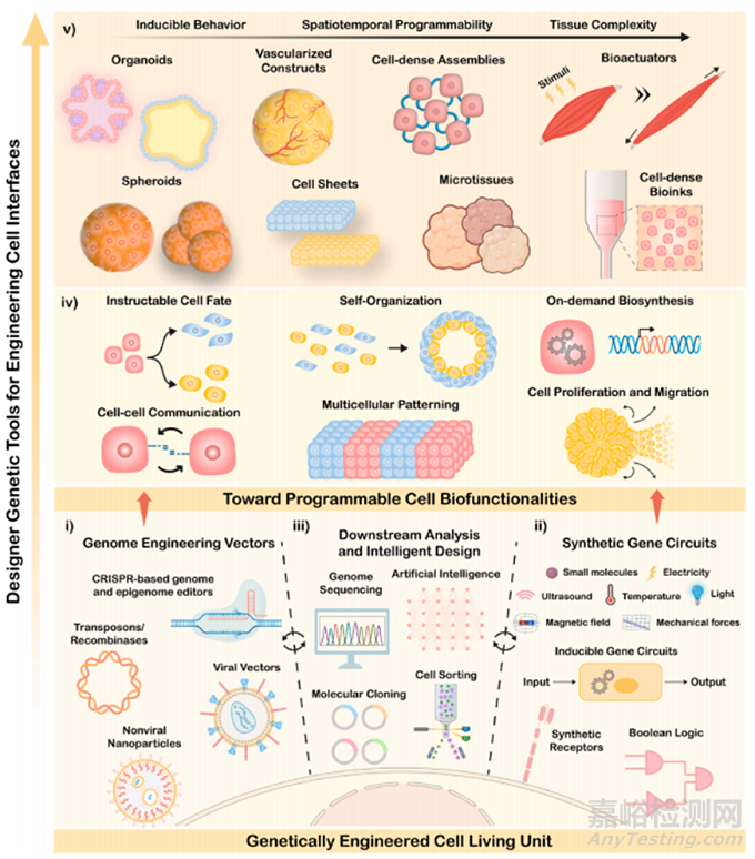
Fig. 1. Illustration of genetically programming mammalian cell living building blocks for bioengineering complex living systems. (i) Inside-out cell engineering strategies include major transposon and viral vectors, as well as site-specific CRISPR-based genome and epigenome editors, in which proficient delivery systems are key for efficiently delivering such apparatus into the cells. (ii) Inducible synthetic biology-inspired gene circuits are key for controlling cell behavior. This can be attained by exploiting designed synthetic receptors, closed-loop circuits responsive to exogenous stimuli (e.g., light, small molecules, sound), and open-loop feedback circuits responsive to specific physiological cues. Logic gated circuits are also key for achieving cellular biocomputing programmability. (iii) In parallel, downstream analysis and intelligent design approaches aid in the efficient and rapid development of such engineering toolboxes, harnessing emergent artificial intelligence algorithms, next-generation genome sequencing, cell sorting strategies, and molecular cloning. (iv) At a cellular level, different living behaviors can be meticulously controlled, including on-demand synthesis of specific biomolecules, cell fate and differentiation, proliferation and migration, as well as cell-cell communication and gene expression patterning. (v) For fabricating truly living mammalian systems, inside-out engineering can be interfaced with outside-in engineering approaches (e. g., materials science, biofabrication and tissue engineering), for developing complex designer constructs with high biofunctionality and spatiotemporal programmability. Organoids, spheroids, vascularized constructs, cell sheets, programmable cell-dense assemblies and microtissues, dynamic bioactuators and bioinks can be engineered for human therapeutics, regenerative medicine, disease modelling, and soft robotics.
3、推进基因定制哺乳动物活体材料
基因工程哺乳动物活材料最近出现在广泛的生物医学应用中,将组织工程平台提升到具有定义程序的复杂生物构建体。通过探索此类工具,可以从内到外对细胞进行编程,以随着时间的推移调整其行为,例如允许研究人员:(i)塑造组装的生命结构的架构和功能和/或(ii)编码生命疗法中的智能特征,如将在以下子章节中展示的。
3.1塑造多单元组件的架构和功能
在合成组织发育和形态发生方面,有机体可被视为三维自组织实体。这些实体一般可以来自人类胚胎干细胞(hESC)、诱导多能干细胞(hiPSC)或成人干细胞(hASC)。这类生物微组织可真实再现人体组织的早期发育状态或功能,也可作为独特的体外疾病建模平台。为了克服生物工程方面的挑战,从根本上构建越来越多的生物仿真类器官组织,近年来出现了各种基因引导的类器官组织(图 2)。早期的尝试是利用转录因子 GATA6 的多克隆诱导表达来组装复杂的胎儿肝脏类器官,其表型可再现人的芽和血管样。
在一种优雅的方法中,探索了信息计算转录组学分析,通过过度表达关键转录因子(即 ATF5、PROX1)和基于 CRISPR 的特定酶(即细胞色素 P450 3A4 (CYP3A4)),合理设计出具有优良肝脏特征和血管性的成熟结构。值得注意的是,血管结构在器官的器官发生、命运成熟和模式化过程中起着关键作用 。通过诱导转录因子 ETV2 的表达,研究人员还能从基因上引导 hESCs 衍生的大脑和hiPSCs 衍生的肾脏器官组织中血管网络的建立。在后者中,广泛内皮化的产生是驱动具有人源化细胞池的类似本地多线肾脏成熟的关键,由此产生的器官组织中含有成熟的荚膜细胞、间质细胞和肾素细胞(图 2A)。还可以探索合成类似于组织器的信号中心,以对器官组织的自组织进行编程。通过化学方法诱导胚状体(EBs)中长程信号Wnt/β-catenin的极化激活,或脑组织器官中Sonic Hedgehog(SHH)通路的极化激活,可以模拟体内早期组织发育过程,获得图案化的拓扑结构。另外,光遗传学电路已被用于时空协调具有仿生人类神经发育特征的 SHH 模式和极化脑有机体(图 2B)。通过将光诱导遗传模块与单细胞和空间转录组学相结合,研究人员可以确定此类构建体的模式特征。如前所述,精确引导这种三维组织变形的能力反过来又有助于在哺乳动物系统中塑造复杂的结构。作为概念验证,研究人员发现了一种利用光遗传学可逆折叠神经器官组织的方法,即利用脊椎动物形态发生过程顶端收缩的关键调节因子 Shroom3。例如,可以实现多种三维组织变形,包括增厚、扁平和管腔收缩,再现哺乳动物的三维动态形态发生过程(图 2C )。最后,可以利用最先进的基因编辑工具箱,通过准确再现与某些疾病相关的特定基因组突变或基因变异,生成先进的等基因体外人类疾病模型。例如,目前已开发出基于电穿孔碱基和质粒编辑生成同种异源 ASC 肝细胞、子宫内膜和结肠类器官系的熟练而多用途的方案。
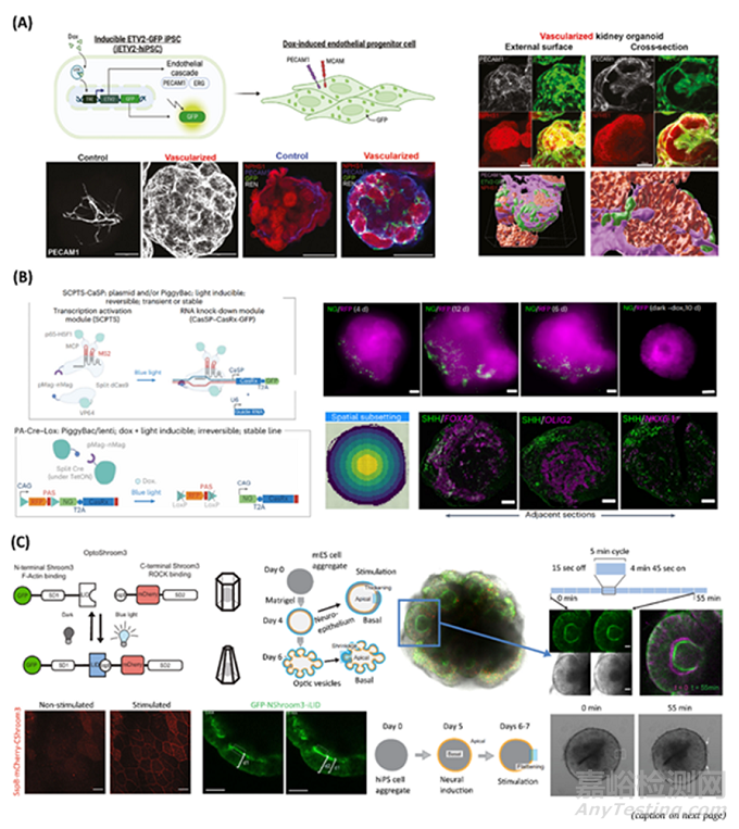
Fig. 2. Strategies and tools for genetically guiding the self-organization and maturation of next-generation organoids. A) Inducible, vascularized human kidney organoid. Top left: Schematic of PiggyBac-engineered dox-inducible ETV2-human induced pluripotent stem cell line (iETV2-hiPSC). Bottom Left: Representative immunofluorescent platelet and endothelial cell adhesion molecule 1 (PECAM-1) images of endothelial cell network in vascularized and control kidney organoids; Images showing emergence of a renin (REN) cell population in vascularized kidney organoid, and no REN+ cells in control. Right: Vascularized kidney organoids with GFP+ endothelial cells encasing the podocyte clusters from the external surface. Reproduced with permission. Copyright 2024, Elsevier. B) Optogenetic patterning of a human neural organoid. Top left: Schematics of light-inducible transcription activation module (SCPTS), based on a dCas9 fused to transcription activation domains, driving CasRx transcription; A U6 promoter-driven CasRx guide RNA can be co-expressed. Bottom left: Schematics of PiggyBac-engineered dox and light-inducible Cre-Lox system, based on a split Cre fused with pMag-nMag photodimers. Top right: Spatial photostimulations by a LED array, in which images represent an organoid (4–12 days), locally photostimulated via laser scanning. Bottom right: Optogenetic stimulation of SHH in neural organoids, coupled with spatial readouts, in which images represent SHH-expressing cells in adjacent cryo-sections of organoids with laser induction of SHH in the north-west pole. Reproduced with permission. Copyright 2023, Springer Nature. C) Optogenetic control of apical constriction in a human neural organoid for induced tissue deformation. Top left: OptoShroom3 genetic constructs, based on dimerization of Shroom Domain 1 (SD1) and Shroom Domain 2 (SD2) upon blue light illumination; Schematics of formation and stages of mouse optic vesicle organoids regarding OptoShroom3-induced tissue deformation. Bottom left: Images of Madin-Darby Canine Kidney (MDCK) cells expressing SspB-mCherry-CShroom3 before and after 1 min stimulation; Images of organoid thickness before and after stimulation. Top right: Optic vesicle organoid and stimulation cycles, altering the lumen diameter of optic vesicles. Bottom right: Images of OptoShroom3-induced flattening through local stimulation of neuroectodermal organoids. Reproduced with permission. Copyright 2022, Springer Nature.
除了干细胞衍生的自组织系统,人工遗传程序也可用于自下而上构建预编程的合成多细胞组合(图3)。受在胚胎发育过程中发现的图灵模式(如斑马皮肤条纹)的启发,可以探索反应扩散数学模型来塑造哺乳动物的模式(即诱导斑点或条纹等)。通过操纵 Nodal-Lefty 信号通路(胚胎形态形成的关键),可以重建激活剂-抑制剂基因回路,诱导短程 Nodal 激活剂的正反馈和长程 Lefty 抑制剂的负反馈。此外,细胞粘附分子(如粘附素)也可以通过策略性操作来引导基于细胞粘附的模式。例如,基于粘附素的细胞分拣系统的化学遗传回路可在带有不同表达的粘附素类型的细胞聚集体中诱导出二维和三维条纹图案。最近,人们探索了一种 synNotch 受体系统,该系统以并列信号受体 Notch 的核心调节结构域为基础,与一个嵌合的细胞外识别结构域和一个嵌合的细胞内转录结构域相连接,从而推动了合成哺乳动物源对称>非对称组织样结构的产生(图 3A)。因此,synNotch 能够生成定制的遗传程序,通过细胞间通信和合成信号级联控制粘连蛋白的表达,从而实现多层图案化小鼠成纤维细胞衍生球体的生物工程(图 3A)。通过细胞信号交互和诱导形态重排的循环逻辑序列,可以实现多细胞自组织的迭代改进。
synNotch 的可重现性和可定制性证明了它在自下而上组织工程学方面的巨大潜力,后来它又被重新设计用于检测可溶性合成形态发生因子。在另一种方法中,为了增加复杂的结构,设计了用于编程图案形成的 helixCAM 平台。该系统基于表面呈现的与跨膜结构域融合的卷曲肽,能够在人类白血病细胞中制造出不同的多细胞聚集体,包括五层球形集合体,并能操纵细胞的迁移和形态。此外,Lim 实验室最近还通过将正交的胞外结合结构域与原生的胞内结构域相结合,生成了新型合成细胞粘附分子(synCAMs)(图 3B)。在这一策略中,synCAMs 可用于决定细胞形态和细胞骨架结构以及可定制的细胞-细胞模式。此外,这些工具还可用于组织重塑:(i) 将不同的细胞群整合到单一的球形结构多细胞结构中;(ii) 使成纤维细胞球体和上皮单层之间自我重塑和融合成复杂的格子状组织网络(图 3B)。未来,这种基因编程结构可加速组织工程和修复的复杂平台。此外,基因回路还可用于在单一细胞类型中精确编程不同的细胞状态。利用锌指转录因子,MultiFate 平台可在外部小分子的调控下不可逆地引导多达七种不同的细胞状态。此外,还探索了一种随机重组酶基因开关,用于从单克隆群体中诱导具有可调亚群比例和三维形态的下游命运。最后,还出现了不同的光遗传学电路,用于将细胞构件组装成组织。例如,E-cadherin 光平台可对 E-cadherin介导的细胞-细胞粘附和肌动蛋白细胞骨架组织进行时空远程编程,从而引导多细胞聚集和迁移(图 3C)。有趣的是,重复的开-关(暗/蓝光)切换周期可用于促进对细胞组装的动态和可逆操作。此类技术可加速研究具有复杂程度的自下而上的多细胞集合体,使哺乳动物生命系统更具活力和仿真性。
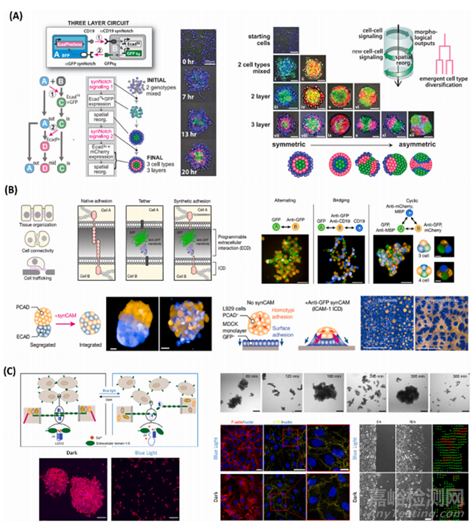
Fig. 3. Bottom-up genetic programming of synthetic multicellular assemblies. A) synNotch-based programming of cell assemblies. Left: Schematics of three-layer circuit, in which an A-type cell sends signals to a B-type cell using CD19 ligand, inducing high expression of E-cadherin and GFP; Induced B-type cell then sends reciprocal signals to A-type cell, and GFP serves as ligand to stimulate anti-GFP synNotch receptor expressed in A-type cell; In the bottom, cell fate diagram showing how the process evolves, self-organizing into three distinct cell phenotypes organized into three spatially distinct compartments; Images of spheroids development with a three-layer architecture, from 0 to 20 h. Right: Representative schematics of different self-organizing multicellular structures programmed via synNotch toolsets. Reproduced with permission. Copyright 2018, American Association for the Advancement of Science (AAAS). B) synCAM-based multicellular assembly and tissue remodeling. Top left: Schematics of functional roles of cell adhesion and design of synCAM receptors, in which the extracellular domain of a CAM is replaced by GFP and a GFP-binding nanobody (anti-GFP). Top right: Custom heterotypic assemblies with alternating, bridging, and cyclic patterning, with L929 cells expressing synCAMs. Bottom left: Exploitation of synCAMs technology to force the integration of differentially sorting L29 populations, leading into a binodal structure. Bottom right: L929 cells mixed with an epithelial MDCK monolayer, initially forming spheroids on top of the monolayer, and later converging into a complex lattice-like network, after the introduction of GFP-anti-GFP synCAMs, thus reshaping tissue organization. Reproduced with permission. Copyright 2022, Springer Nature. C) Opto-E-cadherin-based reversible programming of cell-cell adhesions. Top left: Schematics of Opto-E-cadherin platform, in which cells that express opto-E-cadherin on their surfaces form cell-cell adhesions in the dark but not with blue light; LOV2 domain is inserted between the first and second extracellular domains E-cadherin in proximity to one of the calcium binding sites. In the dark, the Jα-helix of the LOV2 remains folded such that the Ca2+ ions can bind and the E-cadherins on neighboring cells interact. Under blue light, the Jα-helix unfolds such that the Ca2+ ions cannot bind and the E-cadherin interactions are lost. Bottom left: Images showing Opto-E-cadherin-MDA cells after 4 h in the dark (aggregated) or under blue light. Top left: Brightfield images of opto-E-cadherin-MDA cells in suspension culture under repeated 60 min dark/blue light cycles and their clustering dynamics, showing temporal and bidirectional control over cell-cell adhesions. Bottom middle:Fluorescence images showing light-controlled actin cytoskeleton reorganization in the dark or under blue light in 2D cultures, with F-actin (red), nuclei (blue), and p120 (yellow) staining. Bottom right: Bright field images of opto-E-cad-MDA cells in a wound healing assay in the dark and under blue light for 16 h. Reproduced with permission. Copyright 2022, Springer Nature.
合成生物学与材料科学的融合确实可以推动组装出越来越多的可扩展活体结构(图 4)。例如,synNotch 技术最近已升级为一种用途广泛的材料-细胞可编程工具,可通过用户定义的基因表达模式和细胞命运,在空间上控制组织结构。不同的生物材料,如细胞产生的细胞外基质蛋白和水凝胶,都可以用合成配体(如 GFP 和 mCherry)来设计(图 4A)。此外,用 GFP 和 mCherry 配体双重图案化的微流体基底还能使双系受体成纤维细胞选择性地同时共转分化为成肌细胞系和内皮细胞系。这种双重分化可在具有定制组织模式几何形状和排列的连续组织结构中诱导,而无需添加可溶性分化因子。未来,这种技术可与细胞密集型生物墨水相结合,用于构建三维生物打印大尺度图案构建体,甚至用于自我协调器官组织的成熟。值得注意的是,三维生物打印技术已经开始与设计细胞相结合,以实现对活体结构的复杂控制。在最近的一项研究中,利用由多克隆诱导转录因子 hiPSCs 组成的生物墨水组装多细胞神经组织结构。这种方法可使用户定义的神经干细胞、内皮细胞和神经元在错综复杂的分层结构中共同分化,模拟原生组织。从更高的角度来看,对生物打印组合体进行远程时空遗传控制可能特别有趣。在这方面,最近通过将嵌入式挤压体积打印与光遗传工程细胞相结合,开发出了厘米级的细胞密集合成微凝胶。
这种小尺度组织具有光触发胰岛素分泌的胰腺β样细胞的指定模式,展示了高功能微凝胶。此外,通过在具有可灌注通道网络的生物打印水凝胶中装载热诱导细胞,研究人员设计出了 HEAT 平台--用于启动转录的热交换器(图 4B)。从本质上讲,可以利用这些平台实现对三维打印人工组织中体积热诱导基因图谱的深度和规模灵活控制,并具有空间和时间可调性。这种策略对未来组织工程和再生医学的应用可能很有意义。在其他方法中,基因程序被进一步用于组装类似机器的活体材料,这些材料具有驱动、类似生命的运动或甚至非自然的特征,有可能推动具有用户编程功能的软生物混合机器人新时代的到来。在过去几年中,人们设计了多种光遗传学电路,用于在坚固的合成结构中控制肌肉组织收缩。例如,这种电路可以远程编程骨骼生物机器人的多向行走能力,以及功能神经肌肉单元内的鞭毛动态游泳运动。这些进展表明,编程生命材料有望实现日益智能化的生命系统。展望未来,新型细胞-细胞信号可编程工具箱在多能细胞系中的应用有可能实现真正精细的全新类器官平台,并具有定制的模式化和细胞命运决定功能。在这方面,Fussenegger 实验室的开创性工作可作为未来的主要基础,在细胞疗法方面,从疾病反馈闭环到音乐和意念控制的基因调谐,先进的基因回路取得了众多发展。
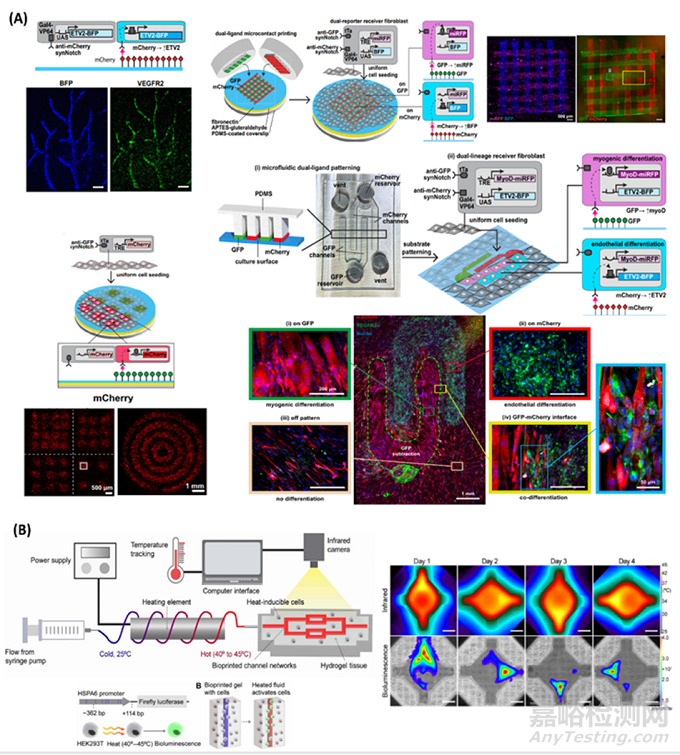
Fig. 4. Upscaling genetically programmed and patterned living cell assemblies. A) Material-to-cell synNotch-based programming of multicellular constructs. Top left: Schematics of receiver fibroblasts with anti-mCherry/Gal4 synNotch that activates BFP and ETV2 when cultured on substrates with mCherry, promoting endothelial differentiation; Vasculature-like pattern with resulting fluorescence microscopy images after three days. Top right: Schematics of dual-ligand microcontact printing and seeding of dual-receiver fibroblasts with anti-GFP/tTa synNotch, activating miRFP and anti-mCherry/Gal4-VP64 synNotch that activates BFP; Fluorescence images of microcontact-printed perpendicular lines of GFP an mCherry and corresponding miRFP and BFP expression by dual-receiver cells, after one day. Bottom left: Schematics of anti-GFP/tTA synNotch receiver fibroblasts that activate mCherry cultured on substrates microcontact-printed with GFP; mCherry fluorescence images of patterns, expressed by receiver cells cultured on GFP-patterned substrates, after two days. Bottom right: Schematics of microfluidic patterning platform with alternating rows of GFP and mCherry and corresponding microfluidic device, for promoting spatial co-differentiation of dual-lineage receiver cells into myogenic and endothelial lineages; Dual-lineage receiver fibroblasts with anti-GFP/tTA synNotch that activates MyoD and miRFP and orthogonal anti-mCherry/ Gal4-VP64 synNotch that activates ETV2 and BFP, cultured on corresponding substrates patterned with GFP and mCherry; In the bottom, image of dual-lineage receiver cells cultured on the patterned substrates, after three days. Reproduced with permission. Copyright 2024, Springer Nature. B) Heat-inducible regulation of gene expression in artificial tissues. Top left: Schematics of HEAT (heat exchangers for actuation of transcription) platform, in which a biocompatible fluid flows around a power supplied heating element to preheat the fluid before entry in perfusable channel networks within hydrogel tissue constructs laden with heat-sensitive cells. During heating, the hydrogel temperature is continuously monitored using an infrared camera. Bottom left: Schematics of HEK293T cells engineered to express luciferase (fLuc) under HSPA6 promoter. Right: Representative infrared and bioluminescence expression images of dynamic hydrogel activation in different days, showing different activated gene expression patterns through space and time. Reproduced with permission. Copyright 2020, American Association for the Advancement of Science (AAAS)
3.2为精准治疗编码更智能的活体生物植入物
为了在活体材料设计中充分应用基因工程工具,可以对细胞进行更智能化的编码,以提高其治疗潜力。随着目前以细胞为基础的疗法取得广泛成功,由哺乳动物细胞组成的基因编程活体组合可能会在下一代个性化干预方面取得前所未有的治疗效果。值得注意的是,在过去的几年中,已经建立了许多合成基因回路,以确保针对各种人类疾病(从癌症到自身免疫或代谢紊乱)的细胞疗法越来越强大。
受这些进展的启发,早期的尝试主要集中在基因升级细胞片、球体或支架支持的组合。例如,通过利用关键转录因子的过度表达,或在多细胞组合中敲入治疗基因盒,实现了基因增强组织再生或伤口愈合干预。尽管如此,在生物相关组织结构中加入合成生物学启发设计的细胞,有望加快治疗性活系统的发展(图 5)。Guilak 实验室在预编程智能生物植入物方面取得了多项进展,这些植入物可自主响应生物线索,具有类似机器的动态行为。例如,利用软骨组织中的机械感应通道 TRPV4,将水凝胶生物植入物与软骨细胞结合在一起,形成机械回路。在生理机械负荷下,这种设计的构建体可以按需感知并自我调节 IL-1 受体拮抗剂(IL-1Ra)的释放,从而保护组织免受促炎症条件的影响。在另一种方法中,CRISPR/Cas9 编辑的 iPSCs 被用于生物工程活软骨组织,并在三维编织支架(图 5A)和琼脂糖棒状软骨植入物中组装自我调节的抗细胞因子疗法。从本质上讲,通过感知炎症性 IL-1,此类生物系统可以利用可诱导的巨噬细胞趋化蛋白-1(Ccl2)启动子,以反馈方式对生理水平的 IL-1Ra 做出反应。
未来,这种闭环人工植入物可在不同的人类疾病条件下进行探索,以实现自主、稳健的细胞给药平台。此外,远红光控制的免疫调节设计细胞(FLICs)已被载入水凝胶,以实现对癌症靶向免疫治疗干预的远程无痕控制(图 5B)。在按需照射时,这种生物植入物可释放 INF-β、TNF-β 和 IL-12 细胞因子,促进抗肿瘤术后复发的长期活性,最终延长动物的存活率。将复杂的电气和软件原理引入哺乳动物的生命系统,可以进一步增加关键层的复杂性和功能性。作为概念验证,已开发出用于糖尿病治疗的智能手机调控光遗传水凝胶结构(图 5C)。在这种平台中,用户控制的智能手机可以远程激活水凝胶中的发光二极管,进而诱导光遗传工程细胞释放人胰高血糖素样肽的短变体(shGLP-1)或胰岛素。在同一系统中,还可进一步利用蓝牙激活的血糖仪实时测量体内生理血糖水平,以无线闭环网络的方式自动触发技术级联。
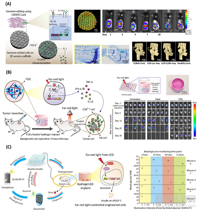
Fig. 5. Bioengineering smarter therapeutic mammalian living materials. A) Gene edited self-regulated anti-cytokine bioimplant for treatment of inflammatory diseases. Left: Schematics of CRISPR/Cas9-based iPSCs engineering with a synthetic gene circuit expressing IL-1Ra, in response to activation of the Ccl2 promoter. Cells were loaded on 3D cartilaginous woven constructs in chondrogenic media. Topcenter: Gene edited cells on porous 3D woven scaffold (nano-computedtomography). Top right: Longevity of implanted constructs, demonstrated by consistent luciferase expression, in mice. Bottom center: Implants reduced inflammation. Bottom right: Mice treated with Ccl2/IL-1Ra implants demonstrate reduced bone damage. Reproduced with permission. Copyright 2022, American Association for the Advancement of Science (AAAS). B) Optogenetic immunotherapeutic construct for cancer treatment. Left: Schematics of optogenetic perioperative immunotherapy mediated by far-red light-controlled immunomodulatory engineered cells (FLICs), encapsulated within a polysaccharide-based hydrogel. Top right: Mixture of far-red light-controlled immunomodulatory designer cells (FLICs) with hydrogel polysaccharide solution, promoting crosslinking of hydrogel matrix, through crosslinking by ions interactions from the cell culture medium and solidified. Bottom right: In vivo bioluminescence imaging of tumor recurrence, being significantly reduced only under far-red light (FRL) illumination, with an LED array (λ=730 nm, 1 mW/cm2 ). Reproduced with permission. Copyright 2022, Springer Nature. C) Smartphone-controlled optogenetic implant for treatment of diabetes. Left: Schematics of smartphone-optogenetically regulated electronic system. Smartphone-remote controlled field generator of HydrogelLED implants induce expression of insulin of shGLP-1. Signals of blood glucose are sent via bluetooth to electronic system for autonomously activating the therapeutic system. Right: LED intensity activating designer cells, as reported on the smartphone. Reproduced with permission. Copyright 2017, American Association for the Advancement of Science (AAAS).
总的来说,我们之前描述了基因工程哺乳动物活体材料和应用的一些主要例子,以及其制造中采用的由内而外的策略和细胞类型,并在下表1中进行了总结。
4. 展望及未来展望
设计具有基因编码功能的哺乳动物活体材料的工作进展迅速,前景广阔。这种由内而外的技术已经得到了明智的探索,使工程组织具有本地发现和用户可控的活体特征(即血管化、图案化、适应性、驱动等),从而全面提高了它们在疾病建模、组织工程和软机器人等应用领域的探索能力。
基因组和表观基因组编辑器正在快速发展,无疤痕大型转基因插入的新兴平台,如TwinPE、PASTE、PASSIGE或STRAIGHT-IN,可以预见这些可以使越来越复杂的合成基因盒在哺乳动物细胞中的熟练整合。
此外,设计“底盘 ”细胞具有更复杂的可切换行为、新型合成受体和信号通路,可加速生成可定制的活体结构。电遗传学可使研究人员设计出下一代生物电子活体界面,甚至具有电可调性的整个合成活体组织。更进一步说,CAR-T 细胞的进步也能启发未来的设计,例如利用布尔电路组装逻辑门控智能活体系统,或利用安全开关(如自杀或开/关操作)实现高度可控的细胞行为。我们预计,在这样一个处于早期阶段的领域,未来的进步将以由内而外战略的不断努力为标志。
展望未来,具有材料基因界面开关的结构材料对于在空间上调节细胞命运或嵌入材料的设计细胞的活动非常有意义。在这种情况下,我们设想未来与精密化学、微流体技术和生物制造相关的进步将成为推动细胞密集型活体材料发展的关键。例如,基因编辑的三维生物仿真片上器官技术可以简化更具临床价值的人源化同种疾病模型[7]。此外,在可预见的未来,设计细胞与含有生物传感器的片上器官的结合有可能实现对生物数据的实时监测。
尽管前景广阔,但鉴于这类系统的复杂性,可以预见许多挑战。与工程组织和细胞产品目前在临床转化方面遇到的监管障碍类似,基因赋能的活体材料预计也会在基因递送载体的效率、制造、可重复性、细胞源标准化和安全性方面遇到重大障碍。此外,由于合成生物学可能赋予研究人员编码新颖/非天然特征的能力,伦理方面的影响也可能上升。iPSCs 衍生的治疗组合可从特定基因编辑中获得巨大收益,以减少潜在的免疫原性痕迹。此外,单细胞 CRISPR 筛选等技术也极具优势,可在体外对有机体中的基因调控通路进行高通量分析。未来,一个潜在的挑战可能是如何确保细胞在活体材料中长时间保持功能,这可能会阻碍细胞的最终应用。

来源:Internet